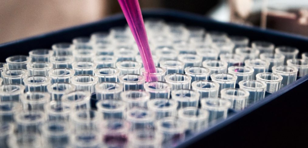
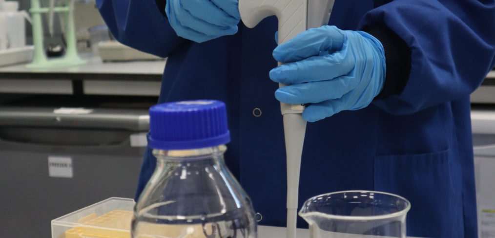
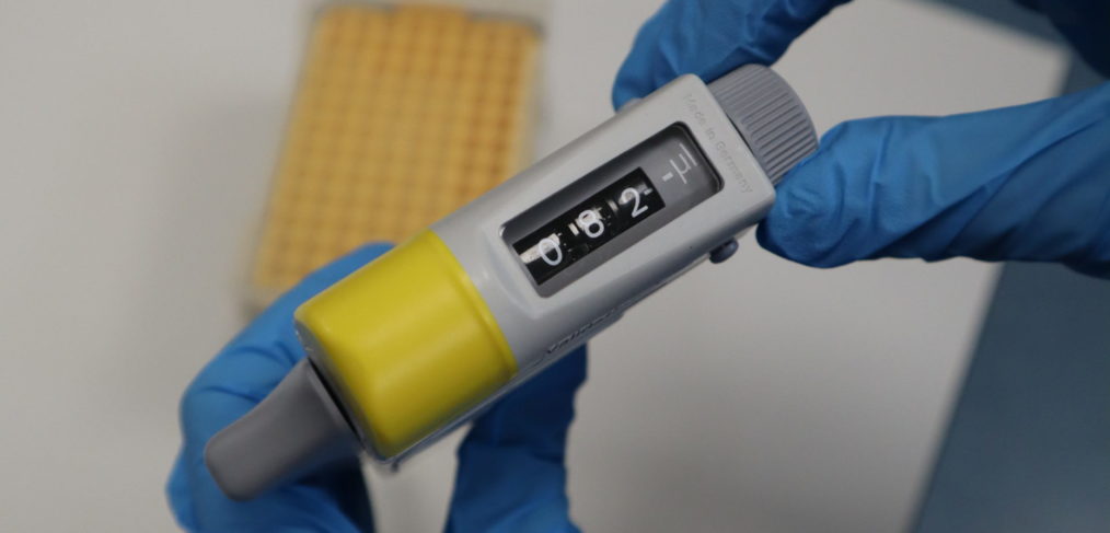
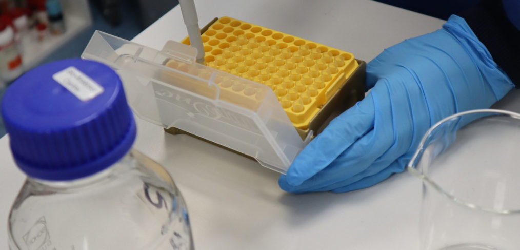
After learning the correct way to pipette Bio Analysis invited me back to continue training. Watch the video to find out how I did and don’t hesitate to contact us about any enquires.
If want to partake in this 1 day course or any other services we provide then please contact us
Simultaneous Analysis of 97 Primary Metabolites on a Pentafluorophenylpropyl Column (PFPP). This assay was performed by Shimadzu (1), the manufacturer of the equipment used, all information shown is from them unless otherwise stated.
What are Metabolites?
‘Metabolites are substances made or used when the body breaks down food, drugs, chemicals, or its own tissue (for example, fat or muscle tissue). This process, called metabolism, makes energy and the materials needed for growth, reproduction, and maintaining health. It also helps get rid of toxic substances’ (2).
Energy at the cellular level is derived by various metabolic processes, including the glycolytic system and the TCA cycle. In order to investigate the metabolites of living systems it is important to quantify the amount of each metabolite.
Method and Sampling
Many primary metabolites are hydrophilic and difficult to analyse by reverse phase chromatography. Usually, Ion pair chromatography is used for hydrophilic compounds, but this is not compatible with LCMS analysis due to the ion pair reagents causing high background signals and sensitivity deterioration.
A method using a PFPP column has been developed to overcome these limitations, it allows not only hydrophobic interaction but also retention of hydrophilic compounds, which is essential for successful analysis of primary metabolites.
The samples provided to Shimadzu were tissue extracts and all measurements were done on a Shimadzu LCMS – 8040 Triple Quadrupole UFMS.
Liver and Heart tissue samples where extracted and immediately frozen in liquid nitrogen, each frozen sample was weighed and homogenised in methanol containing internal standards. After homogenisation, a methanol-chloroform extraction was carried out, the metabolites were collected, and the extracts concentrated.
After preparation, 3 µl of the solution was injected onto the column using the gradient shown in Table 2 at a flow rate of 0.25ml / min.
In each extract over 80 metabolites were detected, the PFPP column was especially effective at separating the amino acids and organic acids (1).
References:
This assay was performed by Shimadzu (1), the manufacturer of the equipment used, all information shown is from them unless otherwise stated.
Mycotoxins are severely toxic contaminants which can be found in grains, they’re naturally occurring and are produced by certain moulds found on food (fungi) and present a risk to human health. The most common are Aflatoxins and Ochratoxin A. The World Health Organisation set the maximum level for Aflatoxin in food at 0.5 – 15 µg/kg (2) and for Ochratoxin-A at 1 – 50 µg/mg for food and 100 – 1000 µg/kg for animal feed (3).
Due to the toxicity of Mycotoxins, the rapid determination of their presence is essential. UHPLC-MS/MS offers the best combination of selectivity, sensitivity, and speed for detecting these compounds in complex matrices. In this study (1), a high throughput method for the quantitation of 23 Mycotoxins in various matrices was established.
Method:
5g of dry food (such as grain or animal feed) was mixed with 10ml of water to produce the sample. 10g of wet food (such as fruit) produced the sample.
10ml Acetonitrile was added and the sample macerated, before salts were added to allow phase separation. The solution was then centrifuged to pellet the solids. The supernatant was transferred to a clean tube and 5 fold diluted with Mobile Phase A (Water + 0.5% acetic acid) and internal standard.
20 µl of the solution was injected onto the column using the gradient shown in Table 1 at a flow rate of 0.4ml / min.
This assay could be used for any type of food, and as the method is developed by the manufacturer on the exact machine, we can run this assay. If you have a similar project would like analysed, then please get in contact.
References
The Royal Veterinary College (RVC) and the Bio-Analysis Centre are now conducting testing for atypical myopathy as part of the RVC’s work towards improved treatments and management of this disorder, and to enhance the welfare of affected horses. Atypical myopathy of horses is a severe and life threatening equine muscle disorder that is caused by the ingestion of Sycamore tree seeds, leaves or seedlings by horses that are kept at pasture. Risk factors for horses remain unclear. It is, for example, not currently known whether some trees are more toxic than others or whether the amount of toxin varies at certain times of the year or with certain climatic conditions. The RVC is working to help horse owners to gain a better understanding of the condition.
Following research that was supported by The Horse Trust and the RVC’s Animal Care Trust (ACT), the Comparative Neuromuscular Diseases Laboratory at the RVC is now offering testing of seeds, seedlings and leaves for the hypoglycin A toxin known to cause this disorder. To find out if plant samples from your property contain the toxin known to cause atypical myopathy, you can now send samples directly to the lab where they will be tested at a subsidised cost of £50. In addition, the Comparative Neuromuscular Diseases Laboratory is also offering testing of horse blood and urine samples, submitted by your vet, if they suspect atypical myopathy or in field companions. This should help to establish a much more rapid and accurate diagnosis, and subsequent treatment, than with previous tests.
Professor Richard Piercy, Professor of Comparative Neuromuscular Disease, said: “We’re really pleased to be able to launch our testing service for owners who may be concerned about their horses. With the support of the Horse Trust and ACT, and through working with owners in this way, we hope to be able to improve the understanding of atypical myopathy and improve the welfare of horses with this severe condition.”
Whilst electrophoresis is the process during which ions undergo movement in a fluid or gel under the influence of an electric field, capillary electrophoresis is a technique that separates these ions based on their electrophoretic mobility with the use of an applied voltage. This mobility is dependent on the atom’s radius, the charge of the molecule, and the molecule’s viscosity. The rate at which the charged particle moves is directly proportional to the applied electric field – as the field strength increases the mobility increases also.
The gadgetry and the skills required, for the successful application of such techniques, are less so common. Having spent the summer months of 2017, working for the Bio Analysis Centre, I have been a very keen observer of the company’s modus operandi. I have been most impressed by the assembly of systems, developed and maintained by the laboratory manager, Dr. Hyde. This framework for the BAC ensures the smooth running of the services it provides.
Our Bio-Analysis Centre has over 20 years experience in LC-MS and HPLC analysis.